
Mouse brain pictures at their sharpest 2023
X-rays can’t see delicate, watery tissue, but MRI can. Even while MRI can detect a brain tumor, it cannot see the brain’s tiny order.
In a decades-long technical feat led by Duke’s Centre for In Vivo Microscopy, researchers improved MRI resolution to capture the sharpest mouse brain images ever.
To celebrate the 50th anniversary of the first MRI, scientists developed mouse brain pictures that are far clearer than clinical MRIs for people. From an 8-bit pixelated picture to a Chuck Close painting is the scientific equivalent.
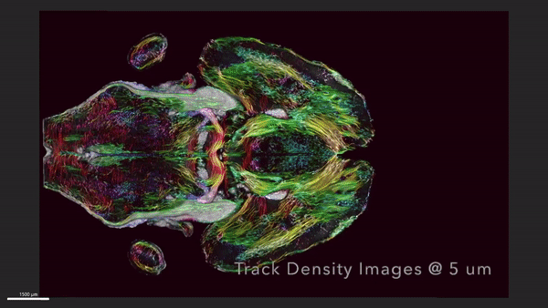
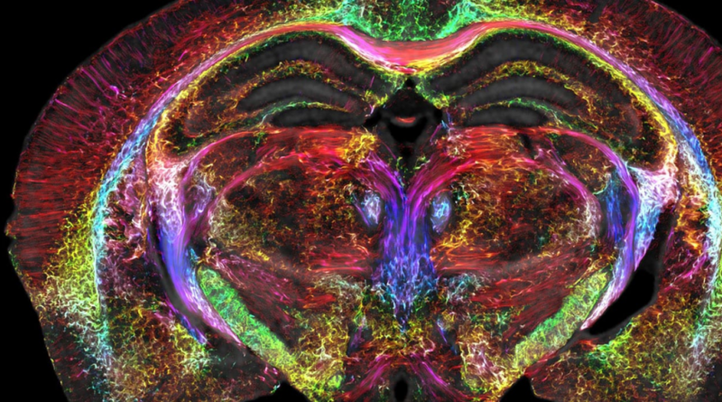
The new pictures’ voxels, or cubic pixels, are 5 microns. Compared to a clinical MRI voxel, it’s 64 million times smaller.
Researchers utilized magnets mostly on mice. The improved MRI allows unprecedented visualization of brain connections. These mice imaging findings may help us understand human situations including how the brain changes with age, food, and neurodegenerative disorders like Alzheimer’s.
“It is truly enabling,” said G. Allan Johnson, Ph.D., the primary author of the new work and Duke’s Charles E. Putman University Distinguished professor of radiology, physics, and biomedical engineering. Neurodegenerative illnesses can be seen differently.”
A powerful magnet (most clinical MRIs use magnets between 1.5 and 3 Tesla; researchers use a 9.4 Tesla magnet), a unique set of gradient coils that are 100 times more powerful than those in a clinical MRI and help create the brain image, and a high-performance computer that is roughly equivalent to nearly 800 laptops working together to image one brain are essential components.
Johnson and his team submit the tissue to light sheet microscopy after “scanning the daylights out of it.” To follow Parkinson’s disease development, they can label dopamine-producing brain cells using this additional technique.
The team then overlays the original MRI scan, which is more anatomically exact and provides a vivid depiction of cells and circuits throughout the brain, with the light sheet images, which are very accurate brain cell imaging.
Thanks to this unified whole-brain data imaging, researchers may now investigate the brain’s smallest secrets.
Researchers can now see the brain’s tiny secrets thanks to whole brain data images.
MRI pictures reveal how brain-wide connectivity changes as mice mature and how certain areas, such the memory-involved subiculum, change more than the rest of the brain.
In a mouse model of Alzheimer’s disease, a spool of rainbow-colored brain connections shows the astonishing neural network degeneration.
By making the MRI a more powerful microscope, Johnson and colleagues aim to better comprehend mice models of human disorders like Huntington’s and Alzheimer’s. And it should help you grasp how comparable things work or go wrong in individuals.
“Research supported by the National Institute of Aging found that modest dietary and drug interventions could lead to animals living 25% longer,” Johnson added. Is their brain intact over this prolonged lifespan? Can they solve crosswords? Will they be able to solve Sudoku despite living 25% longer? It’s now visible. And we can immediately apply that to the human condition.”